Magnetic Resonance - Technology Information Portal Welcome to MRI Technology
Info
  Sheets

Out-
      side


 'Contrast Enhanced Magnetic Resonance Angiography' 
SEARCH FOR    
 
  2 3 5 A B C D E F G H I J K L M N O P Q R S T U V W X Y Z
Result : Searchterm 'Contrast Enhanced Magnetic Resonance Angiography' found in 1 term [] and 13 definitions [], (+ 5 Boolean[] results
previous     6 - 10 (of 19)     next
Result Pages : [1]  [2 3]  [4]
Searchterm 'Contrast Enhanced Magnetic Resonance Angiography' was also found in the following service: 
spacer
News  (1)  
 
Contrast Enhanced MRIInfoSheet: - Contrast Agents - 
Intro, Overview, 
Characteristics, 
Types of, 
etc.MRI Resource Directory:
 - Contrast Enhanced MRI -
 
Contrast enhanced MRI is a commonly used procedure in magnetic resonance imaging. The need to more accurately characterize different types of lesions and to detect all malignant lesions is the main reason for the use of intravenous contrast agents.
Some methods are available to improve the contrast of different tissues. The focus of dynamic contrast enhanced MRI (DCE-MRI) is on contrast kinetics with demands for spatial resolution dependent on the application. DCE-MR imaging is used for diagnosis of cancer (see also liver imaging, abdominal imaging, breast MRI, dynamic scanning) as well as for diagnosis of cardiac infarction (see perfusion imaging, cardiac MRI). Quantitative DCE-MRI requires special data acquisition techniques and analysis software.
Contrast enhanced magnetic resonance angiography (CE-MRA) allows the visualization of vessels and the temporal resolution provides a separation of arteries and veins. These methods share the need for acquisition methods with high temporal and spatial resolution.
Double contrast administration (combined contrast enhanced (CCE) MRI) uses two contrast agents with complementary mechanisms e.g., superparamagnetic iron oxide to darken the background liver and gadolinium to brighten the vessels. A variety of different categories of contrast agents are currently available for clinical use.
Reasons for the use of contrast agents in MRI scans are:
Relaxation characteristics of normal and pathologic tissues are not always different enough to produce obvious differences in signal intensity.
Pathology that is sometimes occult on unenhanced images becomes obvious in the presence of contrast.
Enhancement significantly increases MRI sensitivity.
In addition to improving delineation between normal and abnormal tissues, the pattern of contrast enhancement can improve diagnostic specificity by facilitating characterization of the lesion(s) in question.
Contrast can yield physiologic and functional information in addition to lesion delineation.
Imaging of arteries and veins with contrast enhanced angiography (CE MRA).

Common Indications:
Brain MRI : Preoperative/pretreatment evaluation and postoperative evaluation of brain tumor therapy, CNS infections, noninfectious inflammatory disease and meningeal disease.
Spine MRI : Infection/inflammatory disease, primary tumors, drop metastases, initial evaluation of syrinx, postoperative evaluation of the lumbar spine: disk vs. scar.
Breast MRI : Detection of breast cancer in case of dense breasts, implants, malignant lymph nodes, or scarring after treatment for breast cancer, diagnosis of a suspicious breast lesion in order to avoid biopsy.

For Ultrasound Imaging (USI) see Contrast Enhanced Ultrasound at Medical-Ultrasound-Imaging.com. See also Blood Pool Agents, Myocardial Late Enhancement, Cardiovascular Imaging, Contrast Enhanced MR Venography, Contrast Resolution, Dynamic Scanning, Lung Imaging, Hepatobiliary Contrast Agents, Contrast Medium and MRI Guided Biopsy.
 
Images, Movies, Sliders:
 Delayed Myocardial Contrast Enhancement from Infarct  Open this link in a new window
      

Courtesy of  Robert R. Edelman
 Left Circumflex Ischemia First-pass Contrast Enhancement  Open this link in a new window
      

Courtesy of  Robert R. Edelman

 Normal Lung Gd Perfusion MRI  Open this link in a new window
 MRI of the Brain Stem with Temoral Bone and Auditory System  Open this link in a new window
    
SlidersSliders Overview

 Breast MRI Images T1 Pre - Post Contrast  Open this link in a new window
 
Radiology-tip.comradContrast Enhanced Computed Tomography
spacer
Medical-Ultrasound-Imaging.comContrast Enhanced Ultrasound,  Contrast Enhanced Doppler Imaging
spacer
 
• Related Searches:
    • Liver Imaging
    • Contrast Enhanced Magnetic Resonance Angiography
    • Gadolinium
    • Contraindications
    • 2 Dimensional Acquisition
 
Further Reading:
  Basics:
Optimal k-Space Sampling for Dynamic Contrast-Enhanced MRI with an Application to MR Renography
Thursday, 5 November 2009   by www.ncbi.nlm.nih.gov    
  News & More:
FDA Approves Gadopiclenol for Contrast-Enhanced Magnetic Resonance Imaging
Tuesday, 27 September 2022   by www.pharmacytimes.com    
Effect of gadolinium-based contrast agent on breast diffusion-tensor imaging
Thursday, 6 August 2020   by www.eurekalert.org    
Artificial Intelligence Processes Provide Solutions to Gadolinium Retention Concerns
Thursday, 30 January 2020   by www.itnonline.com    
Accuracy of Unenhanced MRI in the Detection of New Brain Lesions in Multiple Sclerosis
Tuesday, 12 March 2019   by pubs.rsna.org    
The Effects of Breathing Motion on DCE-MRI Images: Phantom Studies Simulating Respiratory Motion to Compare CAIPIRINHA-VIBE, Radial-VIBE, and Conventional VIBE
Tuesday, 7 February 2017   by www.kjronline.org    
Novel Imaging Technique Improves Prostate Cancer Detection
Tuesday, 6 January 2015   by health.ucsd.edu    
New oxygen-enhanced MRI scan 'helps identify most dangerous tumours'
Thursday, 10 December 2015   by www.dailymail.co.uk    
All-organic MRI Contrast Agent Tested In Mice
Monday, 24 September 2012   by cen.acs.org    
A groundbreaking new graphene-based MRI contrast agent
Friday, 8 June 2012   by www.nanowerk.com    
MRI Resources 
Directories - Lung Imaging - Breast MRI - Cochlear Implant - Absorption and Emission - Abdominal Imaging
 
Intera 1.5TPanorama 0.2InfoSheet: - Devices -
Intro, 
Types of Magnets, 
Overview, 
etc.MRI Resource Directory:
 - Devices -
 
www.medical.philips.com/de/products/mri/products/ From Philips Medical Systems;
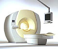
the Intera-family offers with this member a wide range of possibilities, efficiency and a ergonomic and intuitive serving-platform. Also available as Intera CV for cardiac and Intera I/T for interventional MR procedures.
The scanners are also equipped with SENSE technology, which is essential for high-quality contrast enhanced magnetic resonance angiography, interactive cardiac MR and diffusion tensor imaging (DTI) fiber tracking.
The increased accuracy and clarity of MR scans obtained with this technology allow for faster and more accurate diagnosis of potential problems like patient friendliness and expands the breadth of applications including cardiology, oncology and interventional MR.
Device Information and Specification
CLINICAL APPLICATION
Whole body
CONFIGURATION
Short bore compact
Standard: head, body, C1, C3; Optional: Small joint, flex-E, flex-R, endocavitary (L and S), dual TMJ, knee, neck, T/L spine, breast; Optional phased array: Spine, pediatric, 3rd party connector; Optional SENSE coils: Flex-S-M-L, flex body, flex cardiac
Optional
SYNCHRONIZATION
ECG/peripheral: Optional/yes, respiratory gating
PULSE SEQUENCES
SE, Modified-SE (TSE), IR (T1, T2, PD), STIR, FLAIR, SPIR, FFE, T1-FFE, T2-FFE, Balanced FFE, TFE, Balanced TFE, Dynamic, Keyhole, 3D, Multi Chunk 3D, Multi Stack 3D, K Space Shutter, MTC, TSE, Dual IR, DRIVE, EPI, Cine, 2DMSS, DAVE, Mixed Mode; Angiography: PCA, MCA, Inflow MRA, CE
IMAGING MODES
Single Slice 2D , Multi Single Slice 2D, Multi Slice 2D, 3D, Multi Chunk 3D, Multi Stack 3D
TR
2.9 (Omni), 1.6 (Power), 1.6 (Master/Expl) msec
TE
1.0 (Omni), 0.7 (Power), 0.5 (Master/Expl) msec
SINGLE/MULTI SLICE
RapidView Recon. greater than 500 @ 256 Matrix
FOV
Max. 53 cm
0.1 mm(Omni), 0.05 mm (Pwr/Mstr/Expl)
128 x 128, 256 x 256,512 x 512,1024 x 1024 (64 for BOLD img.)
MEASURING MATRIX
Variable in 1% increments
PIXEL INTENSITY
Lum.: 120 cd/m2; contrast: 150:1
Variable (op. param. depend.)
60 cm diameter (patient)
MAGNET WEIGHT
2900 kg
H*W*D
240 x 188 x 157 cm
POWER REQUIREMENTS
380/400 V
CRYOGEN USE
0.03 L/hr helium
STRENGTH
30 mT/m
5-GAUSS FRINGE FIELD
2.4 m / 3.8 m
Passive and dynamic
spacer

• View the DATABASE results for 'Intera 1.5T™' (2).Open this link in a new window

MRI Resources 
Research Labs - Jobs - Jobs pool - MRI Technician and Technologist Jobs - Bioinformatics - General
 
Lung ImagingMRI Resource Directory:
 - Lung Imaging -
 
Lung imaging is furthermore a challenge in MRI because of the predominance of air within the lungs and associated susceptibility issues as well as low signal to noise of the inflated lung parenchyma. Cardiac and respiratory triggered or breath hold sequences allow diagnostic imaging, however a comparable image quality with computed tomography is still difficult to achieve.
Assumptions for lung MRI:
•
Low signal to noise ratio of the inherently low lung proton density.
•
Cardiac and respiratory motion artifacts.
•
Magnetic susceptibility effects of large magnetic field gradients.
•
Very short transverse relaxation times and significant diffusion yielding short T2 (30-70 msec), short T2* (1-3 msec), and additional long T1 relaxation times (1300-1500 msec).
•
The extreme short T2 values are responsible for a fast signal decay during a single shot readout, resulting in blurring.

The current trends in MRI are the use of new imaging technologies and increasingly powerful magnetic fields. Among these technologies are parallel imaging techniques as well as ventilation agents like hyperpolarized helium for the use as an inert inhalational contrast agent to study lung ventilation properties. With hyperpolarized gases clear images of the lungs can be obtained without using a large magnetic field (see also back projection imaging). Single shot sequences (e.g. TSE or Half Fourier Acquisition Single Shot Turbo Spin Echo HASTE) used in lung MR imaging benefits from parallel imaging techniques due to reduced relaxation time effects during the echo train and therefore reduced image blurring as well as reduced motion artifacts.
In the future, more effective contrast agents may provide an alternative solution to the need for high field MRI. Dynamic contrast enhanced MRI perfusion has demonstrated a potential in the diagnosis of pulmonary embolism or to characterize lung cancer and mediastinal tumors. 3D contrast enhanced magnetic resonance angiography of the thoracic vessel.

See also the related poll result: 'MRI will have replaced 50% of x-ray exams by'
 
Images, Movies, Sliders:
 Anatomic Imaging of the Lungs  Open this link in a new window
      

Courtesy of  Robert R. Edelman
 Normal Lung Gd Perfusion MRI  Open this link in a new window
      

Courtesy of  Robert R. Edelman

 MRI Thorax Basal Plane  Open this link in a new window
 
Radiology-tip.comradLung Scintigraphy
spacer

• View the DATABASE results for 'Lung Imaging' (7).Open this link in a new window


• View the NEWS results for 'Lung Imaging' (3).Open this link in a new window.
 
Further Reading:
  Basics:
A safer approach for diagnostic medical imaging
Monday, 29 September 2014   by www.eurekalert.org    
Parallel Lung Imaging(.pdf)
  News & More:
Chest MRI a viable alternative to chest CT in COVID-19 pneumonia follow-up
Monday, 21 September 2020   by www.healthimaging.com    
CT Imaging Features of 2019 Novel Corona virus (2019-nCoV)
Tuesday, 4 February 2020   by pubs.rsna.org    
Polarean Imaging Phase III Trial Results Point to Potential Improvements in Lung Imaging
Wednesday, 29 January 2020   by www.diagnosticimaging.com    
Low Power MRI Helps Image Lungs, Brings Costs Down
Thursday, 10 October 2019   by www.medgadget.com    
Chest MRI Using Multivane-XD, a Novel T2-Weighted Free Breathing MR Sequence
Thursday, 11 July 2019   by www.sciencedirect.co    
Researchers Review Importance of Non-Invasive Imaging in Diagnosis and Management of PAH
Wednesday, 11 March 2015   by lungdiseasenews.com    
New MRI Approach Reveals Bronchiectasis' Key Features Within the Lung
Thursday, 13 November 2014   by lungdiseasenews.com    
MRI techniques improve pulmonary embolism detection
Monday, 19 March 2012   by medicalxpress.com    
  News & More:
Partnership with VIDA to streamline adoption of advanced MRI of the lungs
Monday, 11 September 2023   by www.itnonline.com    
Searchterm 'Contrast Enhanced Magnetic Resonance Angiography' was also found in the following service: 
spacer
News  (1)  
 
Partial Echo
 
(PE) The partial echo technique (also called fractional echo) is used to shorten the minimum echo time. By the acquisition of only a part of k-space data this technique benefits (like all partial Fourier techniques) from the complex conjugate symmetry between the k-space halves (this is called Hermitian symmetry).
The dephasing gradient in the frequency direction is reduced, and the duration of the readout gradient and the data acquisition window are shortened. Partial echo gives a better SNR at a given TE when a smaller FOV or thinner slices are selected, allows a longer sampling time, and a larger water fat shift (WFS, see also bandwidth) due to a lower gradient amplitude. The resolution is not affected. This is often used in gradient echo sequences (e.g. FLASH, Contrast Enhanced Magnetic Resonance Angiography) to reduce the echo time and yields a lower gradient moment. The disadvantage of using a partial echo can be a lower SNR, although this may be partly offset by the reduced echo time.
Also called Fractional Echo, Read Conjugate Symmetry, Single Side View.

See also Partial Fourier Technique and acronyms for 'partial echo' from different manufacturers.
spacer

• View the DATABASE results for 'Partial Echo' (4).Open this link in a new window

 
Further Reading:
  Basics:
Method and apparatus for subterranean formation flow imaging
   by www.google.com    
MRI Resources 
Hospitals - Universities - Implant and Prosthesis - Abdominal Imaging - IR - Sequences
 
Phase Contrast AngiographyMRI Resource Directory:
 - MRA -
 
(PCA) With this method images of the blood flow-velocity (or any other movement of tissue) are produced. The MRI signal contains both amplitude and phase information. The phase information can be used with subtraction of images with and without a velocity encoding gradient. The signal will be directly proportional to the velocity because of the relation between blood flow-velocity and signal intensity.
This is the strength of PCA, complete suppression of stationary tissue (no velocity - no signal), the direct velocity of flow is being imaged, while in TOF (Inflow) angiography, tissue with short T1 (fat or methaemoglobin) might be visualized.
The strength of the gradient determines the sensitivity to flow. It is set by setting the aliasing or encoding velocity (VENC). Unfortunately, phase sensitization can only be acquired along one axis at a time. Therefore, phase contrast angiographic techniques tend to be 4 times slower than TOF techniques with the same matrix.

See also Phase Contrast Sequence, Magnetic Resonance Angiography, Contrast Enhanced Magnetic Resonance Angiography, Flow Effects and Flow Quantification.
 
Images, Movies, Sliders:
 PCA-MRA 3D Brain Venography Colored MIP  Open this link in a new window


spacer

• View the DATABASE results for 'Phase Contrast Angiography' (8).Open this link in a new window

 
Further Reading:
  Basics:
Magnetic resonance angiography: current status and future directions
Wednesday, 9 March 2011   by www.jcmr-online.com    
  News & More:
MR–ANGIOGRAPHY(.pdf)
MRI Resources 
MRI Training Courses - Pathology - Raman Spectroscopy - Shielding - Guidance - Abdominal Imaging
 
previous      6 - 10 (of 19)     next
Result Pages : [1]  [2 3]  [4]
 Random Page
 
Share This Page
FacebookTwitterLinkedIn

MR-TIP    
Community   
User
Pass
Forgot your UserID/Password ?    


Personalized protocols (age, gender, body habitus, etc.) lead to :
more automated planning 
improved patient comfort 
better diagnostics 
optimized image quality 
nothing 

Look
      Ups


MR-TIP.com uses cookies! By browsing MR-TIP.com, you agree to our use of cookies.

Magnetic Resonance - Technology Information Portal
Member of SoftWays' Medical Imaging Group - MR-TIP • Radiology-TIP • Medical-Ultrasound-Imaging • 
Copyright © 2003 - 2024 SoftWays. All rights reserved. [ 29 April 2024]
Terms of Use | Privacy Policy | Advertising
 [last update: 2024-02-26 03:41:00]